Echocardiography, often referred to as an echo, is the diagnostic tool that uses sound waves to create images of the heart. This technology allows medical professionals to see the heart’s structure, function, and blood flow in real-time, enabling them to diagnose a wide range of cardiovascular conditions. Understanding how this technology works and its importance in cardiac care is crucial for both patients and healthcare providers.
An echocardiogram uses high-frequency sound waves, inaudible to the human ear, which are transmitted through a transducer placed on the chest. These sound waves bounce off the heart’s structures, creating echoes that are then processed by a computer to generate detailed images. This non-invasive procedure provides a wealth of information about the heart’s chambers, valves, and surrounding tissues, aiding in the diagnosis and management of various heart conditions. This information is invaluable in determining the best course of treatment for patients.
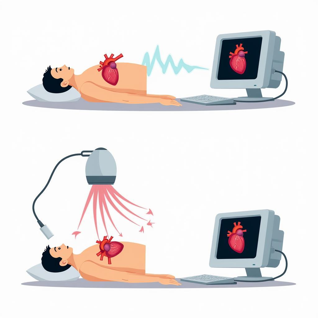 Echocardiography Process
Understanding Echocardiography and its Applications
Echocardiography plays a vital role in diagnosing and monitoring various heart conditions. It can detect issues such as valve abnormalities, heart murmurs, congenital heart defects, and pericardial effusions. Furthermore, echocardiography is essential in assessing the heart’s pumping function, identifying areas of reduced blood flow, and evaluating the effects of treatments.
There are several types of echocardiograms, including transthoracic echocardiography (TTE), transesophageal echocardiography (TEE), and stress echocardiography. Each type has its own advantages and is used in specific situations. For instance, TTE is the most common type and involves placing the transducer on the chest wall, while TEE involves inserting a probe down the esophagus to get a clearer view of the heart. Stress echocardiography is used to assess the heart’s response to exercise or medication.
Different Types of Echocardiograms
Transthoracic Echocardiogram (TTE)
This is the most common type of echocardiogram. A technician places a transducer on your chest, which sends and receives sound waves. TTE can provide information about the size and shape of your heart, the thickness and movement of your heart walls, and the function of your heart valves.
Transesophageal Echocardiogram (TEE)
In a TEE, a small transducer is attached to a thin tube and inserted into your esophagus. This provides clearer images of the heart than TTE, especially of the heart valves.
Stress Echocardiogram
This test involves comparing images of your heart at rest and after exercise or medication that makes your heart beat faster and harder. It helps doctors determine if there are blockages in the arteries that supply blood to your heart.
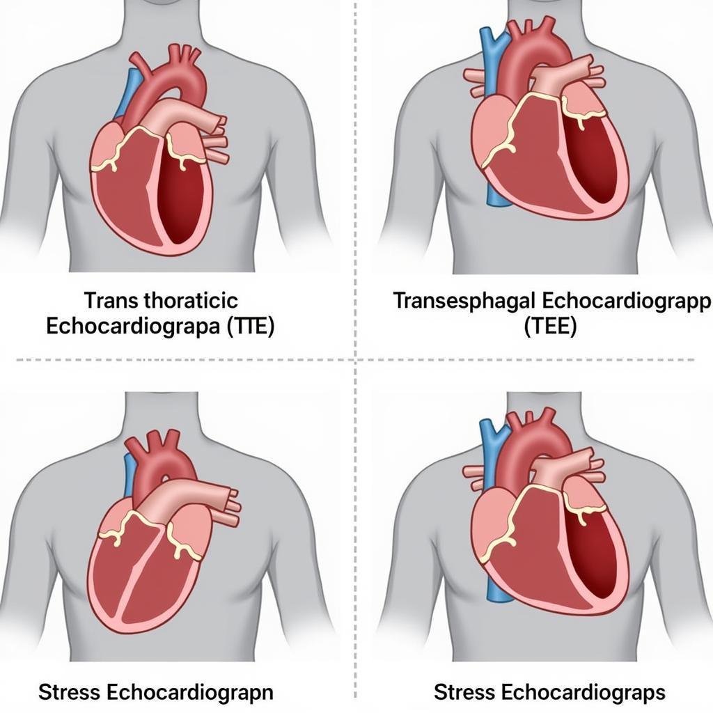 Types of Echocardiograms
The Importance of Early Diagnosis with Echocardiography
Early diagnosis of heart conditions is crucial for effective treatment and improved patient outcomes. Echocardiography, with its ability to provide real-time images of the heart, allows for early detection and timely intervention. This non-invasive and relatively quick procedure can identify potential problems before they become severe, improving the chances of successful treatment and reducing the risk of complications.
“Echocardiography is a cornerstone of cardiovascular diagnostics. Its ability to visualize the heart’s structure and function in real-time is unparalleled,” says Dr. Emily Carter, a leading cardiologist at the University of California, San Francisco.
How to Prepare for an Echocardiogram
Generally, there’s no special preparation needed for a standard echocardiogram. However, for a transesophageal echocardiogram (TEE), you’ll need to fast for several hours before the procedure. Your doctor will give you specific instructions.
 Preparing for an Echocardiogram
The Future of Echocardiography
Technological advancements continue to enhance the capabilities of echocardiography. Three-dimensional echocardiography (3DE) provides even more detailed images of the heart, while newer techniques are improving the accuracy and efficiency of the procedure. These developments are contributing to more precise diagnoses and better patient care.
“The future of echocardiography is bright. New technologies are constantly emerging, allowing us to see the heart in ways we never thought possible,” says Dr. David Lee, a renowned cardiovascular imaging specialist at the Cleveland Clinic.
 Future of Echocardiography
Diagnostic Tools for Heart Failure
For more information about diagnostic tools related to heart conditions, particularly heart failure, you can visit our page on diagnostic tools for heart failure.
In conclusion, echocardiography, using its sound wave technology, is the primary diagnostic tool for visualizing the heart. Its versatility, non-invasive nature, and ability to provide real-time images make it essential for diagnosing and managing a wide range of cardiovascular conditions. Early detection through echocardiography can significantly improve patient outcomes. Contact CARW Workshop at +1 (641) 206-8880 or visit our office at 4 Villa Wy, Shoshoni, Wyoming, United States for more information.
diagnostic tools for heart failure
FAQ
-
Is an echocardiogram painful? No, a standard echocardiogram is painless. You may feel some pressure from the transducer on your chest.
-
How long does an echocardiogram take? It usually takes about 30 to 60 minutes.
-
Are there any risks associated with an echocardiogram? It’s a very safe procedure with minimal risks.
-
What can I expect after an echocardiogram? You can resume your normal activities immediately after the test.
-
When will I get my results? Your doctor will discuss the results with you.
-
How much does an echocardiogram cost? The cost varies depending on your insurance coverage and the facility.
-
What if my echocardiogram shows a problem? Your doctor will discuss treatment options with you based on the results.
